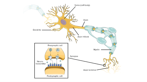
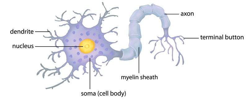
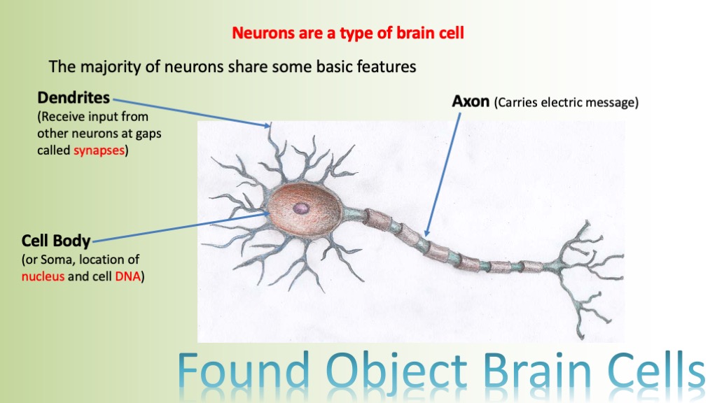
40 art-labeling activity: neuron structure
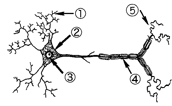
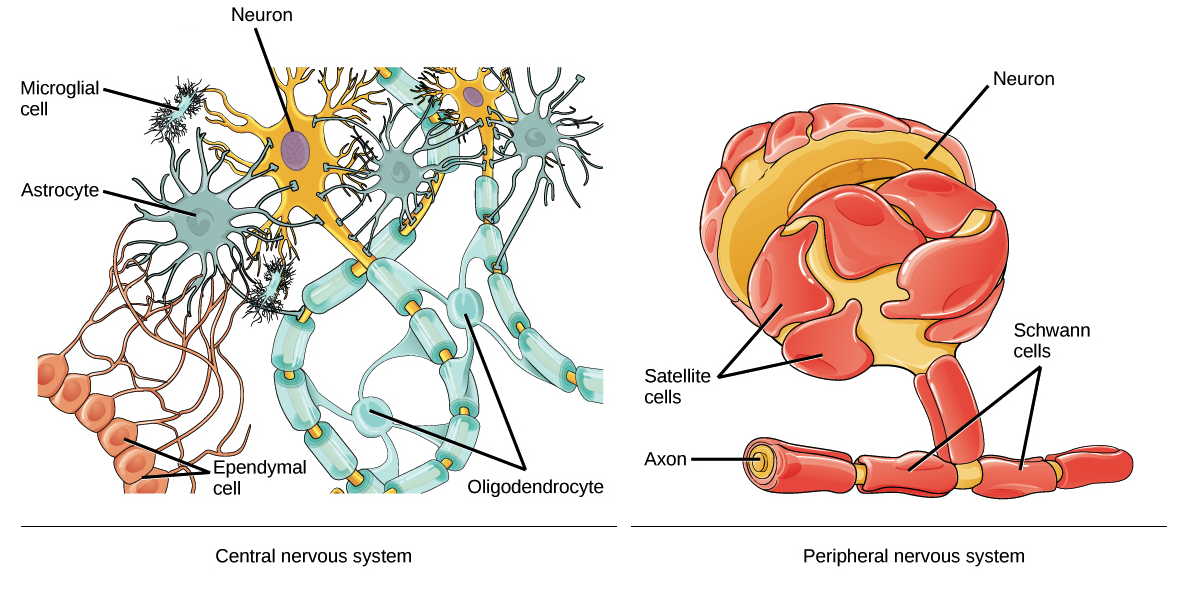
Diagram Quiz on Neuron Structure and Function (Labeling Quiz) Diagram Quiz on DNA replication. 1. Identify the cell type in the above figure. 2. In the figure, labeled '1' receives impulses from adjacent neuron. It is called the. 3. In the figure, labeled '2' is the short filaments from the cell body that carries impulses from dendrites to the cell body which is the. 4. Answer correct art based question chapter 4 question - Course Hero ANSWER: Correct Neuroglial cells have many functions, including anchoring neurons and blood vessels in place, monitoring the composition of the extracellular fluid, speeding up the rate of nerve impulse transmission, and circulating the fluid that surrounds the brain and spinal cord.
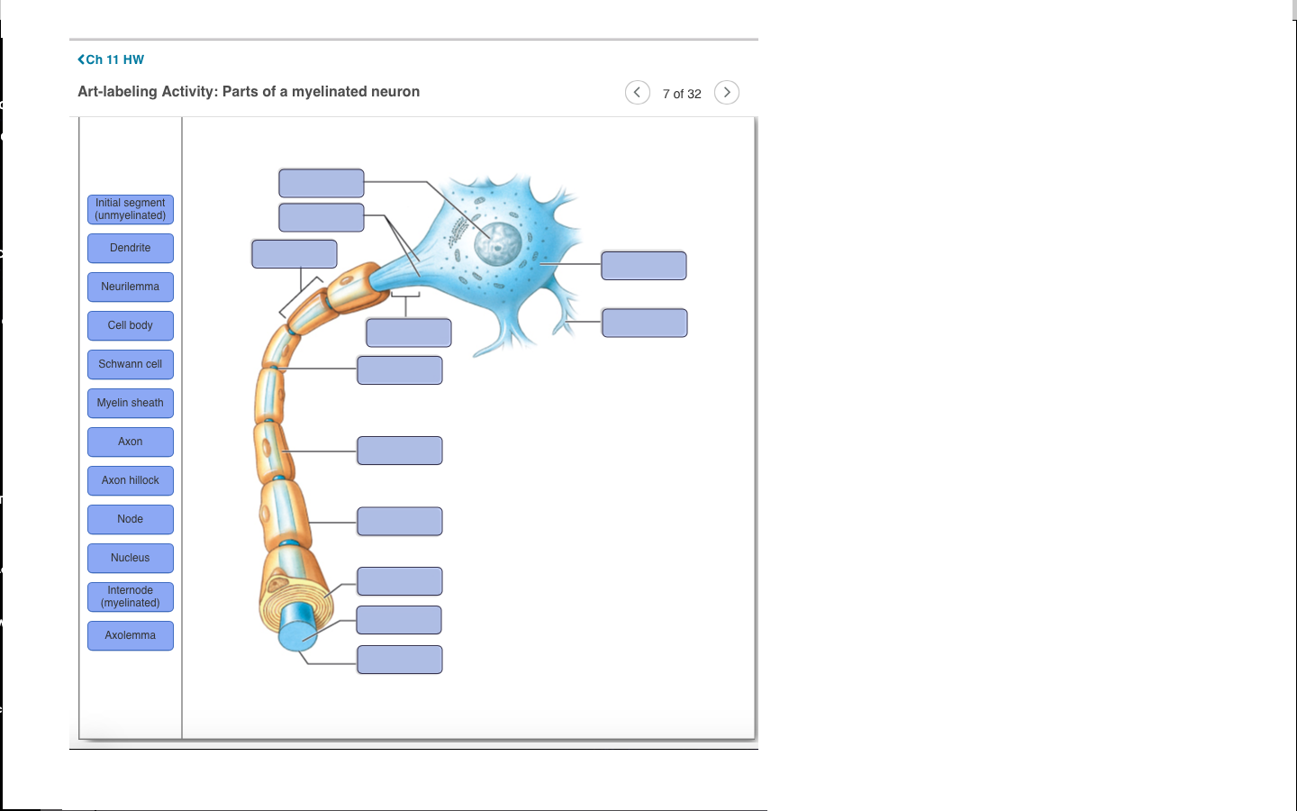
Solved Art-labeling Activity: Parts of a Myelinated | Chegg.com Art-labeling Activity: Parts of a Myelinated Peripheral Nervous System (PNS) Neuron Drag the labels onto the diagram to identify the parts of a myelinated PNS neuron. Reset Help Avon Ilock Nucleos Nodos Donde Initial segment (unmyelinated) Myolinated incomode Axolo Axon Myelin covering internode

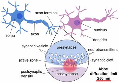
Art-labeling activity: neuron structure
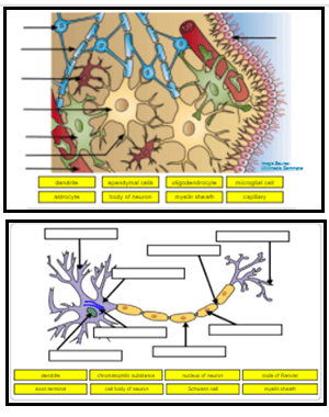
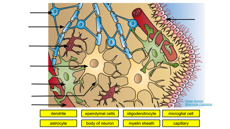
EOF Chapter 12 - Central Nervous System (CNS) Flashcards | Quizlet Art-Labeling Activity: Neuron Structure. Art-Labeling Activity: The Myelin Sheath in the PNS and CNS. Art-Labeling Activity: Neuroglial Cells of the CNS. The image depicts the deterioration of the distal segment of an axon as a result of an injury. This is called _____. Week 4 Chapter 13_.pdf - Course Hero ANSWER: Correct Art-labeling activity: Histology of neural tissue in the CNS A neuron in the cerebrum of the brain sends an impulse to the cerebellum of the brain. A neuron carries information from the stomach area to the brain. A neuron in the spinal cord sends an impulse to the upper limb, removing the hand from a hot object.
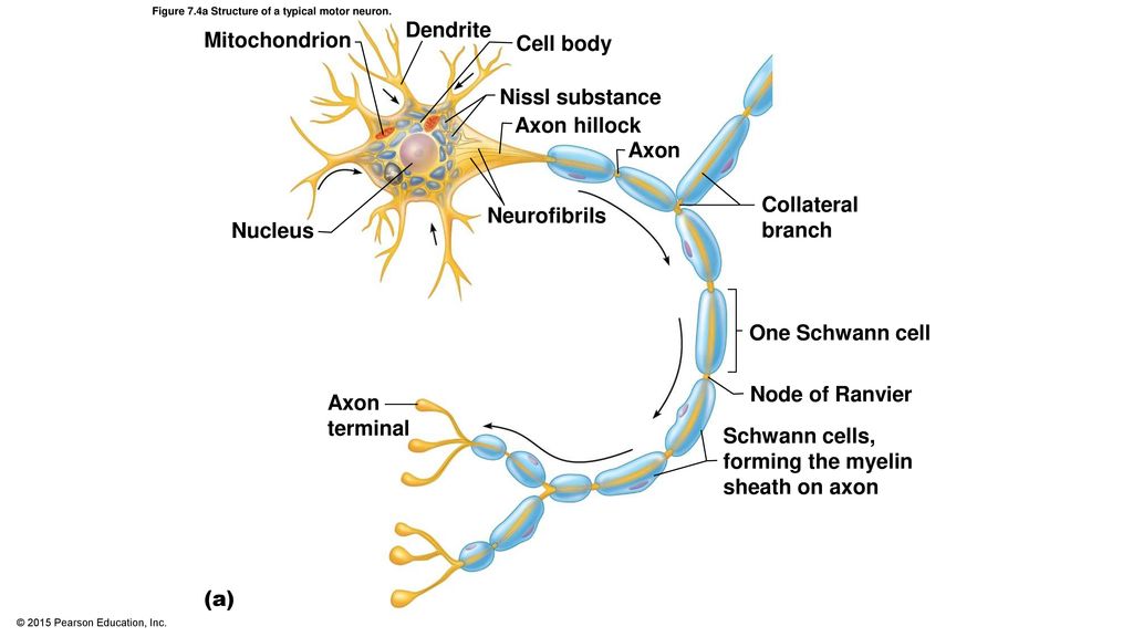
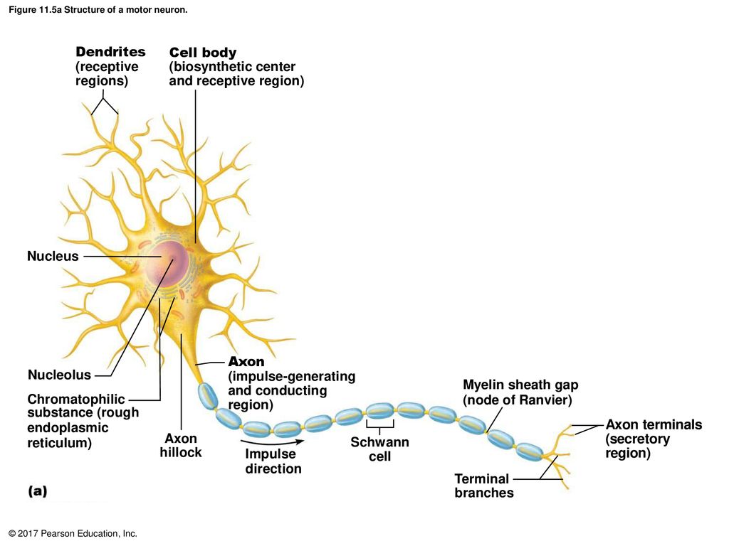
Art-labeling activity: neuron structure. Solved Art-labeling Activity: Neuron Structure 6 of 36 - Chegg Art-labeling Activity: Neuron Structure 6 of 36 Review Part A Drag the labels to the appropriate location in the figure. Axon Nucleus Synaptic terminals Microfibrils and microtubules CO on Coll body II. Mitochondrion Dendrites Nucleolus Submit Request Answer Solv cheo Best chapter 4 adaptive follow-up Flashcards | Quizlet Art-labeling Activity: Neuron structure PICTURE Reading Quiz - Chapter 4 Question 7 Which connective tissue type is found in the walls of large blood vessels and in ligaments supporting transitional epithelia? a) dense irregular connective tissue proper b) elastic connective tissue proper c) adipose connective tissue proper Mastering A&P Chapter 11 - Fundamentals of the Nervous System ... - Quizlet The separation of charges creates a voltage (electrical potential difference), which can be measured using a voltmeter. The resting membrane potential of a neuron averages -70mV (millivolts). All neural activities begin with a change in the resting membrane potential of a neuron. Ch11 HW- Introduction to the Nervous System and Nervous Tissue.pdf Dendrites are branched extensions off of a neuron. Neuroglia are the supporting cells of the nervous system. ... ANSWER: somatic motor division central nervous system peripheral nervous system Correct ArtLabeling Activity: Neuron Structure Part A Drag the appropriate labels to their respective targets.
A&P Ch.11-13 Lecture Test Flashcards | Quizlet Art-Labeling Activity: Neuroglial Cells of the CNS The small phagocytic cells that engulf debris and pathogens in the CNS are the __________. microglia (smallest/ least abundant) The neurotransmitter involved in emotion, motivation, and addictive behavior is __________. dopamine Answered: Art-labeling Activity: Structural… | bartleby Answered: Art-labeling Activity: Structural… | bartleby. Homework help starts here! Science Biology Q&A Library Art-labeling Activity: Structural organization of skeletal muscle Reset Epimysium Muscle fascicle Endomysium Perimysium Nerve Muscle fibers Blood vessels Tendon Muscle fiber (cell) Answered: Art-labeling Activity: Structure of a… | bartleby Art-labeling Activity: Structure of a lymph node Medulla (B cells Cortex (B cells) and macrophages) Medullary sinus Medullary cord Paracortex (T cells) Efferent vessel Capsule Hilum Subcapsular Afferent vessel space Lymph node artery and vein Trabeculae • Previous Question Drag the labels to the appropriate location in the figure. Solved Dendrites Axon Axon hillock Axolemma Myelin sheath - Chegg Art-Labeling Activity: Neuron Structure . there are 4 incorrect and you please labeling it for me. thank you . Show transcribed image text Expert Answer. Who are the experts? Experts are tested by Chegg as specialists in their subject area. We review their content and use your feedback to keep the quality high.
(Get Answer) - Drag the appropriate labels to their ... - Transtutors Drag the appropriate labels to their respective targets. Reset Help Axolemma Axoplasm Dendrites Axon collateral Axon terminals Axon Telodendria Axon Hillock Myelin sheath Cul body A Art-Labeling Activity: Neuron Structure Art-labeling Activity: Special Movements of the Joints 2 Start studying Art-labeling Activity: Special Movements of the Joints 2. Learn vocabulary, terms, and more with flashcards, games, and other study tools. Ch 12 Flashcards | Quizlet Art-labeling Activity: Structure of a typical motor neuron (2 of 2) Which factors contribute to increasing the speed of nerve impulse transmission? larger diameter of axon and the presence of myelin sheath Which of the following statements describes interneurons? Week 4 Chapter 13_.pdf - Course Hero ANSWER: Correct Art-labeling activity: Histology of neural tissue in the CNS A neuron in the cerebrum of the brain sends an impulse to the cerebellum of the brain. A neuron carries information from the stomach area to the brain. A neuron in the spinal cord sends an impulse to the upper limb, removing the hand from a hot object.
Chapter 12 - Central Nervous System (CNS) Flashcards | Quizlet Art-Labeling Activity: Neuron Structure. Art-Labeling Activity: The Myelin Sheath in the PNS and CNS. Art-Labeling Activity: Neuroglial Cells of the CNS. The image depicts the deterioration of the distal segment of an axon as a result of an injury. This is called _____.
EOF















Post a Comment for "40 art-labeling activity: neuron structure"