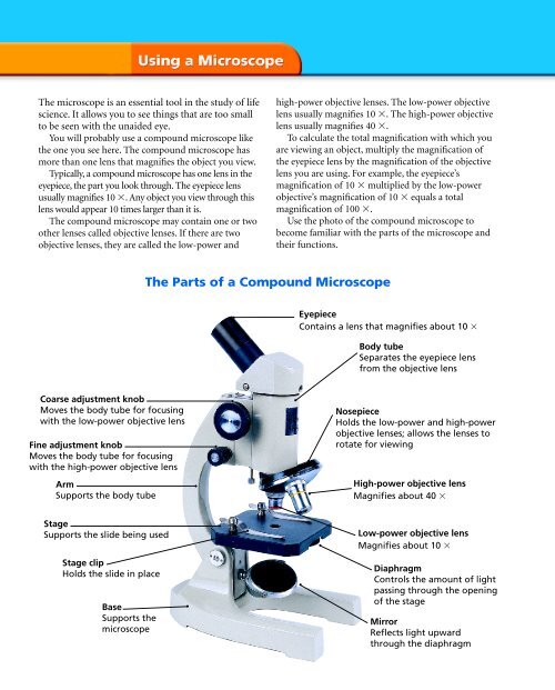
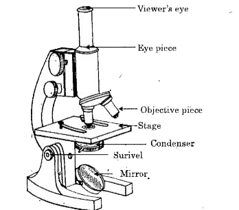
41 compound microscope diagram with labels
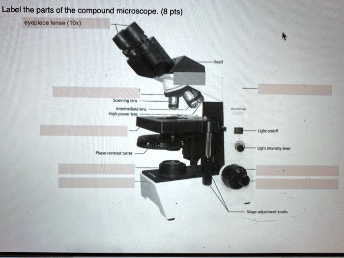
Understanding the Parts of a Compound Microscope: A Detailed Diagram ... #microscope #compoundmicroscope #illustration .A compound microscope diagram typically consists of a series of labeled parts that depict the different compon... Label a Compound Microscope Diagram | Quizlet Start studying Label a Compound Microscope. Learn vocabulary, terms, and more with flashcards, games, and other study tools. ... Label this. Illuminator Switch. Sets found in the same folder. AP II Ch. 24 Digestive Lab QUIZ. 10 terms. CWRN2016. Body planes Label. 9 terms. Hesi_Study.
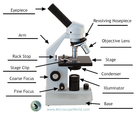
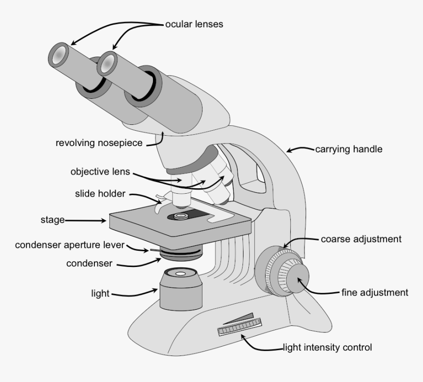
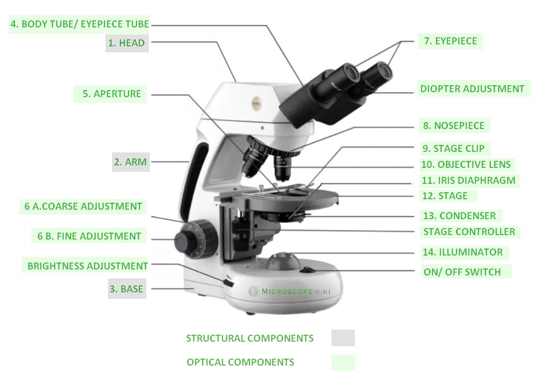
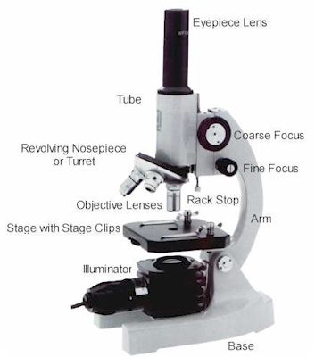
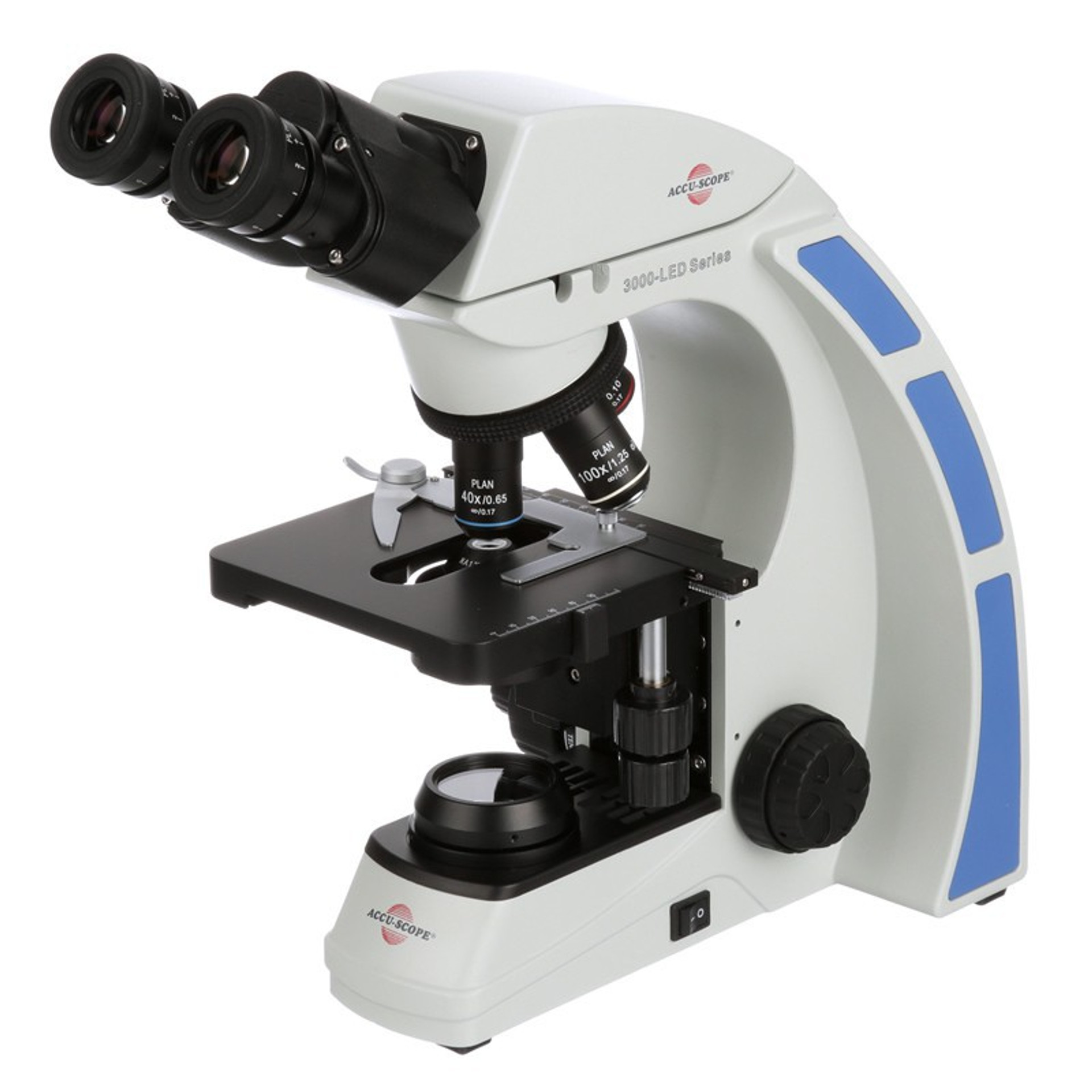
16 Parts of a Compound Microscope: Diagrams and Video The 16 core parts of a compound microscope are: Head (Body) Arm Base Eyepiece Eyepiece tube Objective lenses Revolving Nosepiece (Turret) Rack stop Coarse adjustment knobs Fine adjustment knobs Stage Stage clips Aperture Illuminator Condenser Diaphragm Video: Parts of a compound Microscope with Diagram Explained
Compound microscope diagram with labels
Compound Microscope Parts, Functions, and Labeled Diagram Compound Microscope Definitions for Labels Eyepiece (ocular lens) with or without Pointer: The part that is looked through at the top of the compound microscope. Eyepieces typically have a magnification between 5x & 30x. Monocular or Binocular Head: Structural support that holds & connects the eyepieces to the objective lenses. Microscopy: Intro to microscopes & how they work (article) - Khan Academy In a compound microscope with two lenses, the arrangement of the lenses has an interesting consequence: the orientation of the image you see is flipped in relation to the actual object you're examining. For example, if you were looking at a piece of newsprint with the letter "e" on it, the image you saw through the microscope would be "ə." Label the microscope — Science Learning Hub Label the microscope Interactive Add to collection Use this interactive to identify and label the main parts of a microscope. Drag and drop the text labels onto the microscope diagram. diaphragm or iris base eye piece lens fine focus adjustment light source stage coarse focus adjustment high-power objective Download Exercise
Compound microscope diagram with labels. Compound Microscope: Definition, Diagram, Parts, Uses, Working Principle A compound microscope is defined as A microscope with a high resolution and uses two sets of lenses providing a 2-dimensional image of the sample. The term compound refers to the usage of more than one lens in the microscope. Also, the compound microscope is one of the types of optical microscopes. Parts of a Compound Microscope - Labeled (with diagrams) Parts of a Compound Microscope - Labeled (with diagrams) A compound microscope is known as a high-power microscope that enables you to achieve a high level of magnification. Smaller specimens can be thoroughly viewed using a compound microscope. ... In a compound microscope, the objective lenses are the main lenses which range from 4x to 100x ... Microscope Parts and Functions Microscope Parts and Functions With Labeled Diagram and Functions How does a Compound Microscope Work? Before exploring microscope parts and functions, you should probably understand that the compound light microscope is more complicated than just a microscope with more than one lens. Compound Microscope Parts, Function, & Diagram - Study.com Learn the compound light microscope's parts and functions by viewing a compound microscope diagram. Also, read about the uses of a compound microscope. Updated: 11/04/2021
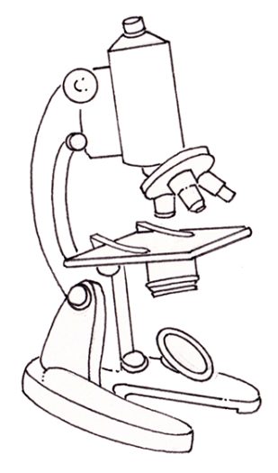
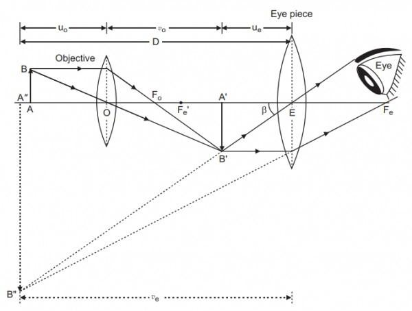
PDF Biological Diagram Of Simple Microscope With Label Labelled Diagram of Compound Microscope Biology Discussion. Fluorescence microscope Wikipedia. 1 1 3 Plant Animal Cell Microscope Lab Wikispaces. Biological Diagram Of Simple ... June 19th, 2018 - To better understand the structure and function of a microscope we need to take a look at the labeled microscope diagrams of the compound and ... Diagram of a Compound Microscope - Biology Discussion Diagram of a Compound Microscope Article Shared by ADVERTISEMENTS: In this article we will discuss about:- 1. Essential Parts of Compound Microscope 2. Magnification of the Image of the Object by Compound Microscope 3. Resolution Power 4. Method for Studying Microbes 5. Measurement of the Size of Objects. Essential Parts of Compound Microscope: Label Parts Of A Compound Microscope Teaching Resources | TPT This is a set of 3 tiered readings. Students will read a passage about the how to use a compound light microscope. Students will use textual evidence to answer questions and label the different parts of the microscope. It also allows students to gain prior knowledge about the compound microscope. Version A provides the most support for students. How to draw compound of Microscope easily - step by step How to draw compound of Microscope easily - step by step Perhaps Bidesh 52.4K subscribers Subscribe 1.4M views 3 years ago Biology diagram I will show you " How to draw compound of microscope...
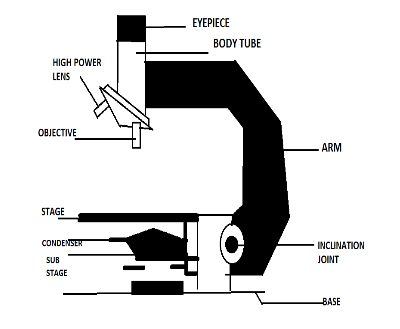
Parts of the Microscope (Labeled Diagrams) - Simple and Compound Microscope Parts of Compound Microscope (Labeled Pictures) a. Mechanical Parts of a Compound Microscope Foot or Base Pillar Arm Stage Inclination Joint Clips Diaphragm Nose piece/Revolving Nosepiece/Turret Body Tube Adjustment Knobs b. Optical Parts of a Compound Microscope Eyepiece lens or Ocular Mirror Objective Lenses Scanning Objective Lens (4x) Compound Microscope Labeled Diagram | Quizlet Compound Microscope Labeled + − Flashcards Learn Test Match Created by meganplocher734 Terms in this set (14) Eyepiece/Ocular lens Contains the ocular lens Body tube A hollow cylinder that holds the eyepiece. Arm Part that supports the microscope. Stage Supports the slide or specimen Coarse adjustment Knob A Study of the Microscope and its Functions With a Labeled Diagram ... These labeled microscope diagrams and the functions of its various parts, attempt to simplify the microscope for you. However, as the saying goes, 'practice makes perfect', here is a blank compound microscope diagram and blank electron microscope diagram to label. Compound Microscope- Definition, Labeled Diagram, Principle, Parts, Uses Compound microscopes have a combination of lenses that enhances both magnifying powers as well as the resolving power. The specimen or object, to be examined is usually mounted on a transparent glass slide and positioned on the specimen stage between the condenser lens and objective lens.
Parts of a microscope with functions and labeled diagram - Microbe Notes Figure: Diagram of parts of a microscope There are three structural parts of the microscope i.e. head, base, and arm. Head - This is also known as the body. It carries the optical parts in the upper part of the microscope. Base - It acts as microscopes support. It also carries microscopic illuminators.
Compound Microscope - Diagram (Parts labelled), Principle and Uses Compound Microscope Parts (Labeled diagram) A compound microscope basically consists of optical and structural components. Within these two systems, there are multiple components within them and they are: Image : Labeled Diagram of compound microscope parts See: Labeled Diagram showing differences between compound and simple microscope parts
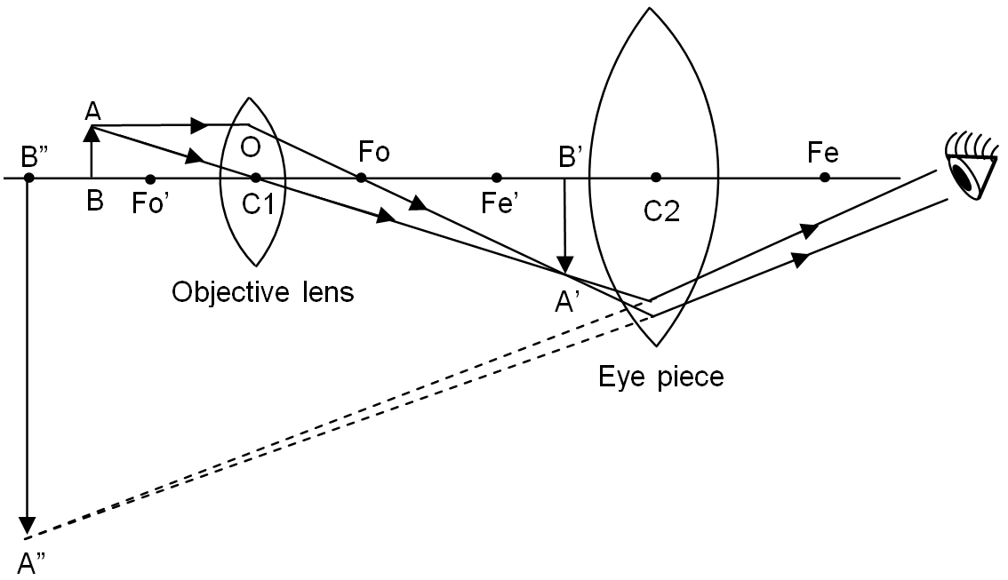
Compound Microscope Labeled Definition, Labeled Diagram, Best procedure ... A compound microscope or compound microscope labeled is a type of microscope that uses two or more lenses to magnify an object. The first lens, called the eyepiece, is used to view the object. The second lens, called the objective lens, is used to collect light from the object and focus it on the eyepiece.
PDF Electron Microscope Diagram Labeled - lindungibumi.bayer.com Electron Microscope Diagram Labeled Electron Microscope Diagram Labeled Diagram Of Electron Microscope Products amp Suppliers. A fluorescence scanning electron microscope ScienceDirect. A tour of the cell Transmission electron microscopy. ... Parts of a Compound Microscope with Diagram and Functions May 8th, 2018 - Before exploring the parts of ...
1.5: Microscopy - Biology LibreTexts In Biology, the compound light microscope is a useful tool for studying small specimens that are not visible to the naked eye. The microscope uses bright light to illuminate through the specimen and provides an inverted image at high magnification and resolution. ... Blank microscope to label parts. This page titled 1.5: Microscopy is shared ...
Compound Microscope Parts - Labeled Diagram and their Functions Labeled diagram of a compound microscope Major structural parts of a compound microscope There are three major structural parts of a compound microscope. The head includes the upper part of the microscope, which houses the most critical optical components, and the eyepiece tube of the microscope.
Label the microscope — Science Learning Hub Label the microscope Interactive Add to collection Use this interactive to identify and label the main parts of a microscope. Drag and drop the text labels onto the microscope diagram. diaphragm or iris base eye piece lens fine focus adjustment light source stage coarse focus adjustment high-power objective Download Exercise
Microscopy: Intro to microscopes & how they work (article) - Khan Academy In a compound microscope with two lenses, the arrangement of the lenses has an interesting consequence: the orientation of the image you see is flipped in relation to the actual object you're examining. For example, if you were looking at a piece of newsprint with the letter "e" on it, the image you saw through the microscope would be "ə."
Compound Microscope Parts, Functions, and Labeled Diagram Compound Microscope Definitions for Labels Eyepiece (ocular lens) with or without Pointer: The part that is looked through at the top of the compound microscope. Eyepieces typically have a magnification between 5x & 30x. Monocular or Binocular Head: Structural support that holds & connects the eyepieces to the objective lenses.












Post a Comment for "41 compound microscope diagram with labels"