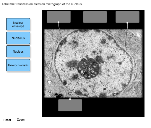
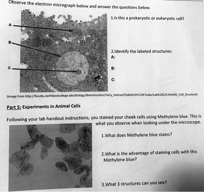
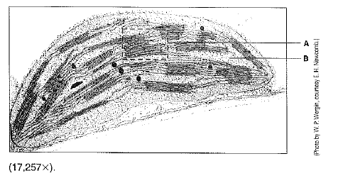
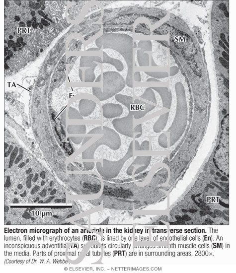
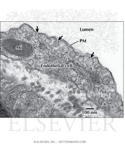
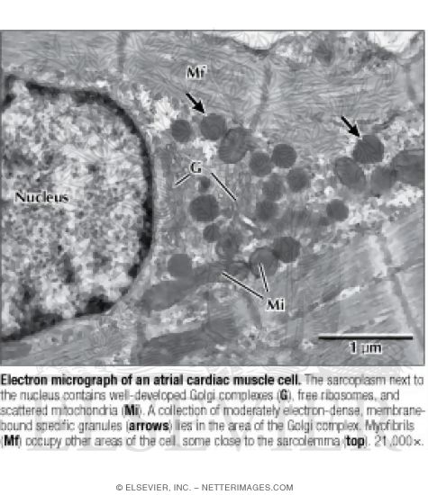
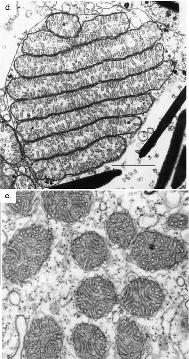
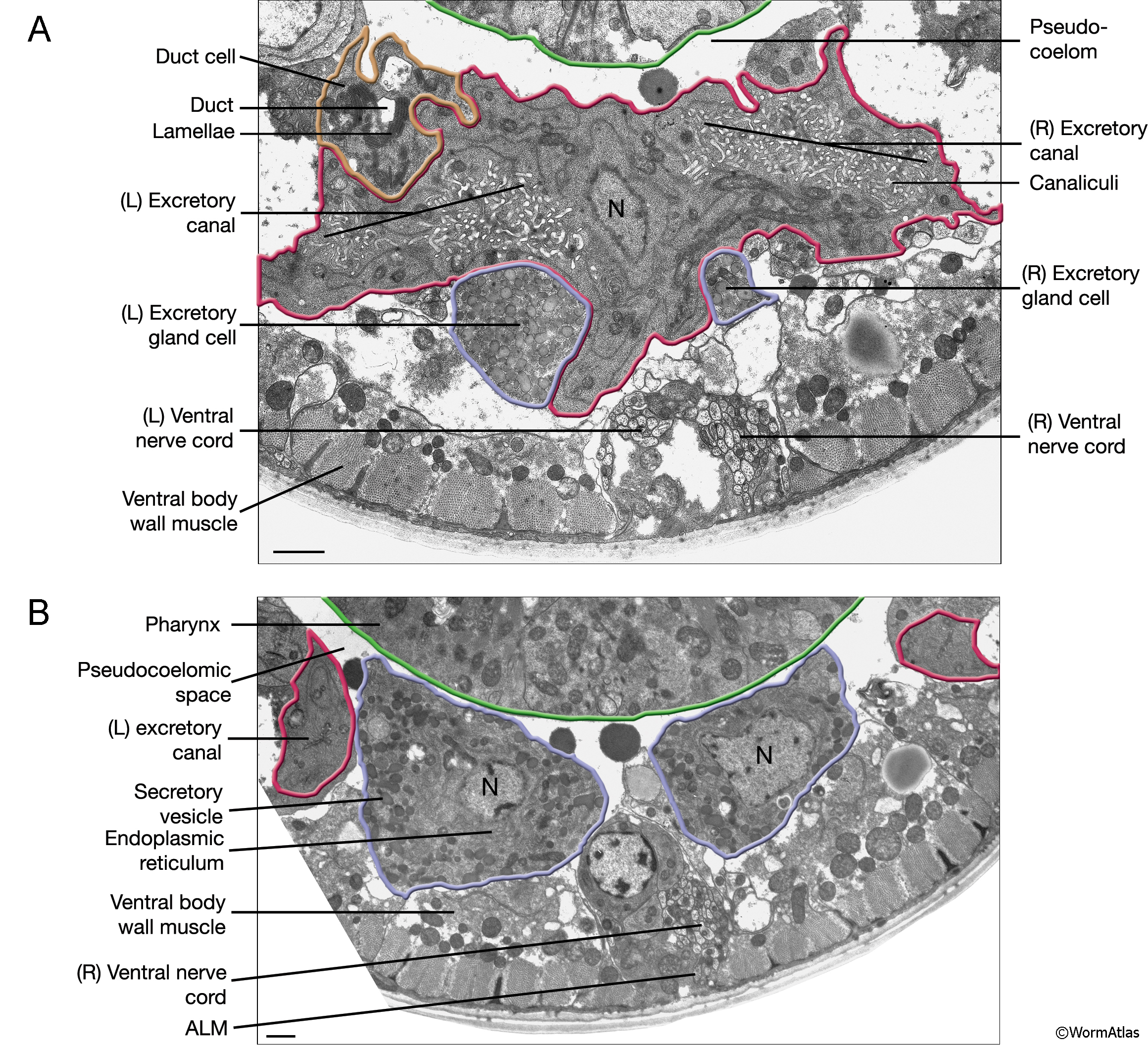
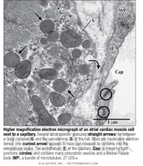
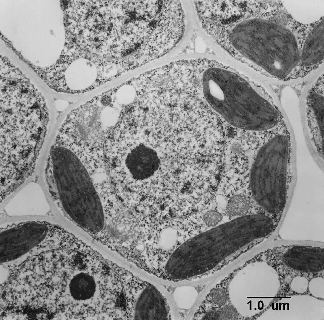
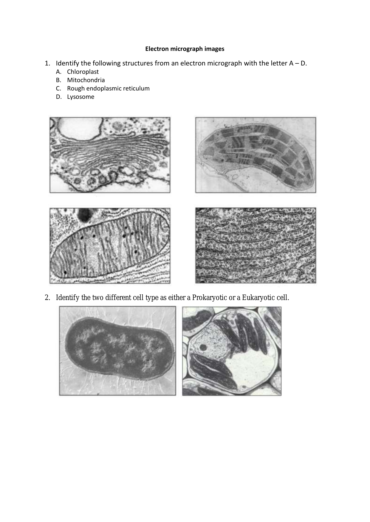
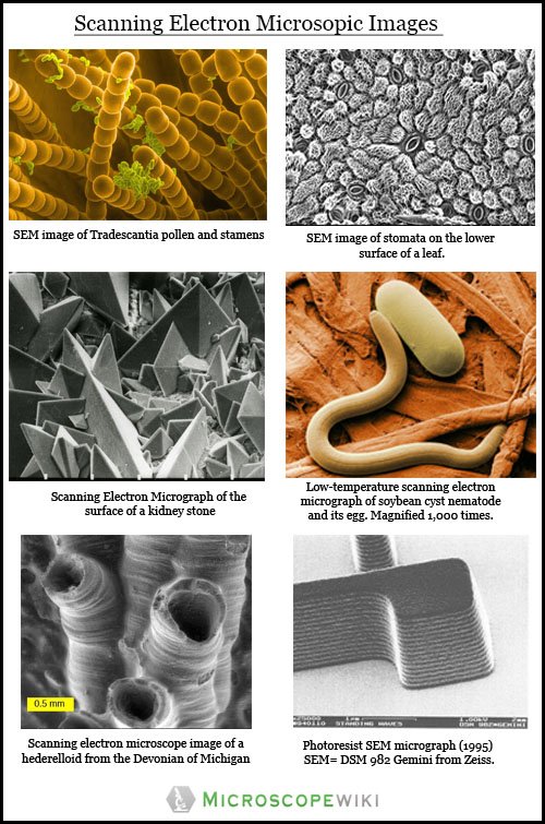
44 electron micrograph labeled
Cell Micrographs - BioNinja Interpretation of electron micrographs to identify organelles and deduce the function of specialised cells. A micrograph is a photo or digital image taken ... Pandemic H1N1/09 virus - Wikipedia WebPandemic H1N1/09 virus Transmission electron micrograph of the pandemic H1N1/09 influenza virus photographed at the CDC Influenza Laboratory. The viruses are 80–120 nanometres in diameter. Virus classification (unranked): Virus: Realm: : …
TEM micrograph of the perinuclear cisternae (pc) in a ... Download scientific diagram | TEM micrograph of the perinuclear cisternae (pc) in a striolar hair cell stained to demonstrate glycogen (Karnovsky, '71). A: The granules in between the cisternae ...

Electron micrograph labeled
Electron micrograph of olfactory bulb from a 2 weeks ... Download scientific diagram | Electron micrograph of olfactory bulb from a 2 weeks manganese-treated mouse, where fibers with areas of myelin disorganization can be seen in the neuropil (Np). Fluorescence microscope - Wikipedia WebA fluorescence microscope is an optical microscope that uses fluorescence instead of, or in addition to, scattering, reflection, and attenuation or absorption, to study the properties of organic or inorganic substances. "Fluorescence microscope" refers to any microscope that uses fluorescence to generate an image, whether it is a simple set up like an … Transmission electron micrograph of the Sq-Ara-C ... Download scientific diagram | Transmission electron micrograph of the Sq-Ara-C nanoaggregates (A). Interaction between [ 3 H]CHE radiolabeled Sq-Ara-C and the different cancer cell lines (B).
Electron micrograph labeled. Parvoviridae - Wikipedia WebGenome. Parvoviruses have linear, single-stranded DNA (ssDNA) genomes that are about 4–6 kilobases (kb) in length. The parvovirus genome typically contains two genes, termed the NS/rep gene and the VP/cap gene. The NS gene encodes the non-structural (NS) protein NS1, which is the replication initiator protein, and the VP gene encodes the viral … Electron microscope - Wikipedia An electron microscope is a microscope that uses a beam of accelerated electrons as a source of illumination. As the wavelength of an electron can be up to 100,000 times shorter than that of visible light photons, electron microscopes have a higher resolving power than light microscopes and can reveal the structure of smaller objects. 1.2 Skill: Interpretation of electron micrographs - YouTube Dec 25, 2015 ... Interpretation of electron micrographs to identify organelles and deduce the functions of specialized cells. Electron micrographs used with ... (A and B) Electron micrograph of a cell labeled for/5-tubulin followed... Download scientific diagram | (A and B) Electron micrograph of a cell labeled for/5-tubulin followed by photooxidation with eosin. The specific staining of ...
Transmission Electron Microscope (TEM)- Definition, Principle, … Web19/05/2022 · Parts of a microscope with functions and labeled diagram; Scanning Electron Microscope (SEM)- Definition, Principle, Parts, Images ; Transmission Electron Microscope (TEM) Images. Transmission electron micrograph of SARS-CoV-2 virus particles, isolated from a patient. Image captured and color-enhanced at the NIAID … AICE Biology Chapter 1: Plant Cell Electron Micrograph Labeling Start studying AICE Biology Chapter 1: Plant Cell Electron Micrograph Labeling. Learn vocabulary, terms, and more with flashcards, games, and other study ... Electron Microscopy of Cells and Tissues | Histology Guide Electron microscopes are widely used to investigate the ultrastructure of biological specimens. Cells and Tissues. Tissues are classified into four basic types: ... Electron Micrographs Below is a collection of electron micrographs with labelled subcellular structures that you should be able to identify. Also, be sure to observe any ...
Site-specific labeling of proteins for electron microscopy - PMC - NCBI Sep 25, 2015 ... The eluted conjugate is now ready for visualization by negative stain or cryo electron microscopy. (D) A representative micrograph containing ... animal cell electron micrograph labelling Diagram - Quizlet Start studying animal cell electron micrograph labelling. Learn vocabulary, terms, and more with flashcards, games, and other study tools. Electron microscope - Wikipedia WebAn electron microscope is a microscope that uses a beam of accelerated electrons as a source of illumination. As the wavelength of an electron can be up to 100,000 times shorter than that of visible light photons, electron microscopes have a higher resolving power than light microscopes and can reveal the structure of smaller objects. A scanning … Transmission electron micrograph of anatase particles from ... Download scientific diagram | Transmission electron micrograph of anatase particles from the ink of the Vinland Map (reproduced from Figure 2 of Towe 1990). Labeled particle sizes are estimated ...
(a) The electron differential flux and the volume emission ... The solid lines correspond to the electron acceleration source with the exponent − 2, the dashed lines corresponds to the exponent 2; (b) same as panel (a) for the heating pulse at 18:54 UT.
Clarifying electron micrograph labeling - JAMA Network Jun 20, 1986 ... Two years ago, a JAMA news article on acquired immunodeficiency syndrome (AIDS) was accompanied by three electron micrographs labeled ...
Virtual EM Micrograph List | histology - University of Michigan Loose Connective Tissue: In this micrograph of loose connective tissue of the tracheal mucosa numerous (labeled) cells of the connective tissue are present. Note the relative size of the different cell types, their shapes, amount of rough ER and variously sized granules and inclusions.
Chloroplast - Wikipedia WebScanning electron micrograph of Gephyrocapsa oceanica, a haptophyte. Haptophytes . Haptophytes are similar and closely related to cryptophytes or heterokontophytes. Their chloroplasts lack a nucleomorph, their thylakoids are in stacks of three, and they synthesize chrysolaminarin sugar, which they store completely outside of the chloroplast, in the …
Integumentary System | histology WebElectron Micrographs. Review Questions. Practice Questions. Suggested Readings. Atlas . Wheater's, pgs. 167-85, Skin . Wheater's, pgs. 95-99 (Glands in general) Wheater's, pgs. 386-90, The breast . Text . Ross and Pawlina (6th ed), Chapter 15 Integumentary System, pgs. 488-525 . Back to Top. Learning Objectives. Be able to identify principal layers of the …
Scanning Electron Microscope (SEM)- Definition, Principle, … Web11/03/2022 · The first Scanning Electron Microscope was initially made by Mafred von Ardenne in 1937 with an aim to surpass the transmission electron Microscope. He used high-resolution power to scan a small raster using a beam of electrons that were focused on the raster. He also aimed at reducing the problems of chromatic aberrations images …
Smallpox - Wikipedia WebThe envelope was labeled as containing scabs from a vaccination and gave scientists at the CDC an opportunity to study the history of smallpox vaccination in the United States. On July 1, 2014, six sealed glass vials of smallpox dated 1954, along with sample vials of other pathogens, were discovered in a cold storage room in an FDA laboratory at the National …
Transmission Electron Microscopy - an overview | ScienceDirect … WebSantwana Padhi, Anindita Behera, in Agri-Waste and Microbes for Production of Sustainable Nanomaterials, 2022. 4.6 Transmission electron microscopy (TEM). Transmission electron microscopy (TEM) is another useful technique of characterization of nanomaterials. It’s a quantitative method to determine the particle size, shape and …
Transmission electron micrograph of the Sq-Ara-C ... Download scientific diagram | Transmission electron micrograph of the Sq-Ara-C nanoaggregates (A). Interaction between [ 3 H]CHE radiolabeled Sq-Ara-C and the different cancer cell lines (B).
Fluorescence microscope - Wikipedia WebA fluorescence microscope is an optical microscope that uses fluorescence instead of, or in addition to, scattering, reflection, and attenuation or absorption, to study the properties of organic or inorganic substances. "Fluorescence microscope" refers to any microscope that uses fluorescence to generate an image, whether it is a simple set up like an …
Electron micrograph of olfactory bulb from a 2 weeks ... Download scientific diagram | Electron micrograph of olfactory bulb from a 2 weeks manganese-treated mouse, where fibers with areas of myelin disorganization can be seen in the neuropil (Np).


















Post a Comment for "44 electron micrograph labeled"