38 diagram of microscope with label
Virtual Labs: Using the Microscope - GameUp - BrainPOP. In this free online science interactive, students learn the procedures for operating a compound optical light microscope as they would use in a science lab. bVX0-zncj9qJ3G1_r18rkIpQL02X-Oi6tWViR4g4-vwDVmU50WZA-4bRZMjM2TXmc88PAkJ1g0jIembnEbM Parts of the Microscope Label and Definition Diagram | Quizlet Medium Power Objective. Provides magnification, usually about 10x; total magnification is 100. High Power Objective. Provides magnification, usually about 40x; total magnification is 400. Stage Clips. Grip slide in place for. viewing. Diaphragm. Controls amount of light entering the body tube.
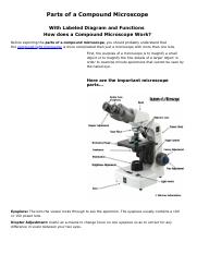
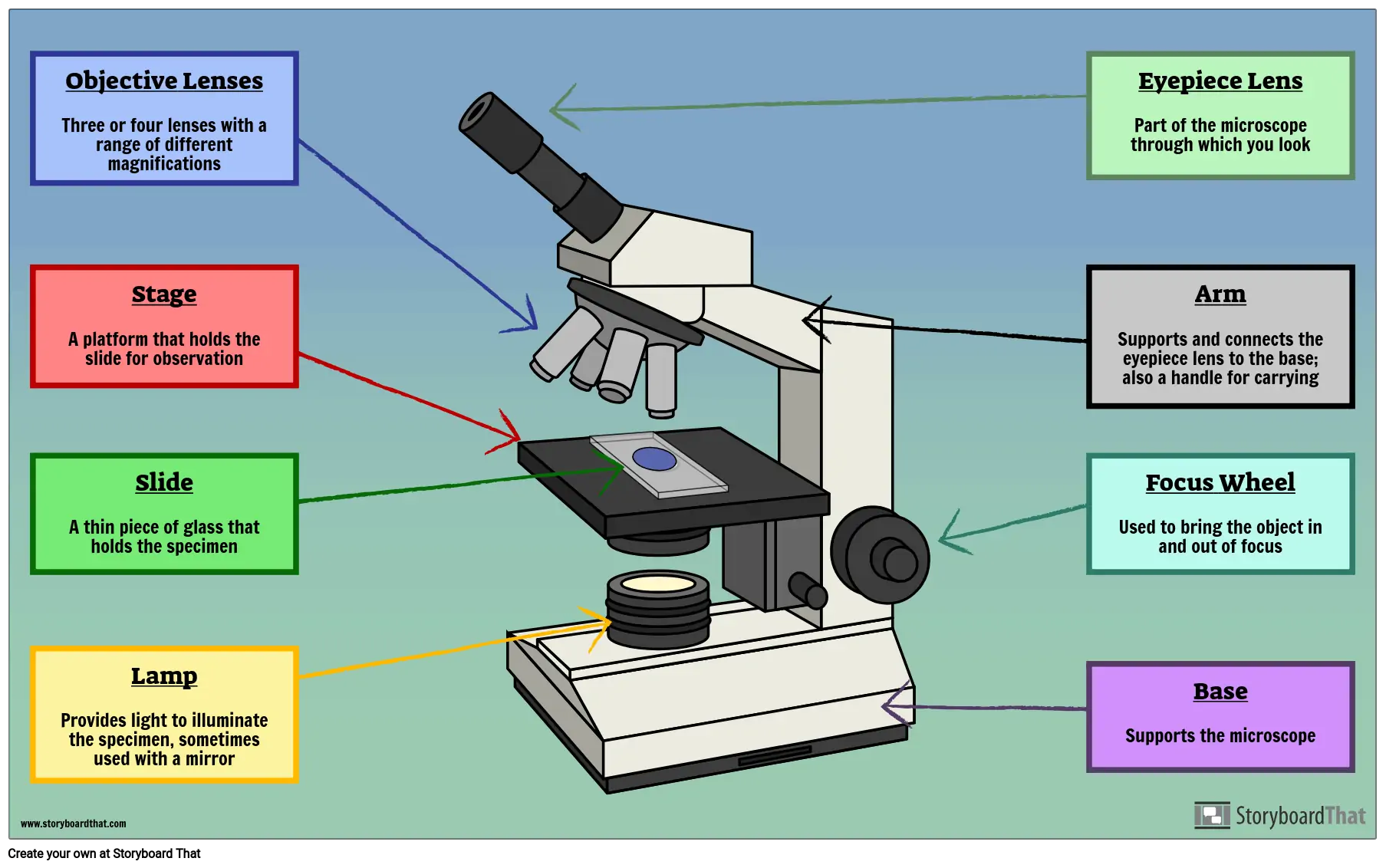
Microscope, Microscope Parts, Labeled Diagram, and Functions 03.09.2022 · Revolving Nosepiece or Turret: Turret is the part of the microscope that holds two or multiple objective lenses and helps to rotate objective lenses and also helps to easily change power. Objective Lenses: Three are 3 or 4 objective lenses on a microscope. The objective lenses almost always consist of 4x, 10x, 40x and 100x powers. The most common eyepiece …

Diagram of microscope with label
Microscope labeled diagram - SlideShare Microscope labeled diagram 1. The Microscope Image courtesy of: Microscopehelp.com Basic rules to using the microscope 1. You should always carry a microscope with two hands, one on the arm and the other under the base. 2. You should always start on the lowest power objective lens and should always leave the microscope on the low power lens ... Parts of Stereo Microscope (Dissecting microscope) – labeled diagram ... Unlike a compound microscope that offers a flat image, stereo microscopes give the viewer a 3-dimensional image that you can see the texture of a larger specimen. [In this image] Examples of Stereo & Dissecting microscopes. Major microscope brands (Zeiss, Olympus, Nikon, Amscope, Omano, Leica …) all produce stereomicroscopes. 16 Parts of a Compound Microscope: Diagrams and Video Once you have an understanding of the parts of the microscope it will be much easier to navigate around and begin observing your specimen, which is the fun part! The 16 core parts of a compound microscope are: Head (Body) Arm. Base. Eyepiece. Eyepiece tube.
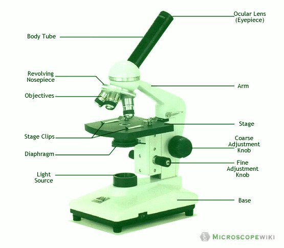
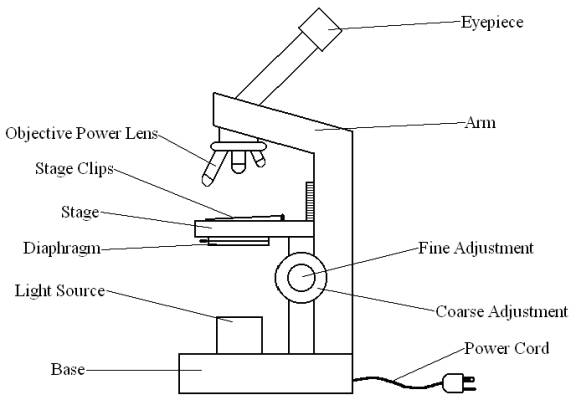
Diagram of microscope with label. Microscope Labeling Diagram | Quizlet Hold the slide in place on the stage. Nosepiece Holds the objective lenses and allows the lenses to rotate for viewing. Stage Supports the slide where the specimen is being viewed. Lamp Projects or reflects light upward through the diaphragm. Base Supports and stabilizes the microscope. Diaphragm Ternary Phase Diagram - an overview | ScienceDirect Topics Ternary phase diagrams are used to represent all possible mixtures of three solvents [1]; they are described in Chapter 3.Here, we shall indicate how they should be used to minimize the solvent consumption. Figure 2.1 (top) shows the methanol–chloroform–water ternary phase diagram with the tie-lines in the biphasic domain. Five particular compositions are shown in the … Simple Microscope - Parts, Functions, Diagram and Labelling Parts of the optical parts are as follows: Mirror - A simple microscope has a plano-convex mirror and its primary function is to focus the surrounding light on the object being examined. Lens - The biconvex lens is placed above the stage and its function is to magnify the size of the object being examined. Label Microscope Diagram - EnchantedLearning.com arm - this attaches the eyepiece and body tube to the base. base - this supports the microscope. body tube - the tube that supports the eyepiece. coarse focus adjustment - a knob that makes large adjustments to the focus. diaphragm - an adjustable opening under the stage, allowing different amounts of light onto the stage.
22 Parts Of a Microscope With Their Function And Labeled Diagram The field diaphragm control is located around the lens located in the base. Hinge Screw -This screw fixes the arm to the base and allow for the tilting of the arm. Stage Clips - They hold the slide firmly onto the stage. On/OFF Switch - This switch on the base of the microscope turns the illuminator off and on. Sperm Under Microscope with Labeled Diagram - AnatomyLearner Sperm under microscope 400x labeled. I will show you the sperm under a microscope 400x with the labeled diagram. Here in the diagram, you will see some seminiferous tubules lined by the thick germinal epithelium. The picture shows the dark Type A and pale Type B spermatogonia located at the seminiferous tubules' basal part. Simple Microscope - Diagram (Parts labelled), Principle, Formula and Uses Parts of a Simple Microscope A simple microscope consists of Optical parts Mechanical parts Labeled Diagram of simple microscope parts Optical parts The optical parts of a simple microscope include Lens Mirror Eyepiece Lens A simple microscope uses biconvex lens to magnify the image of a specimen under focus. Compound Microscope Parts - Labeled Diagram and their Functions Labeled diagram of a compound microscope Major structural parts of a compound microscope There are three major structural parts of a compound microscope. The head includes the upper part of the microscope, which houses the most critical optical components, and the eyepiece tube of the microscope.
Parts of the Microscope with Labeling (also Free Printouts) Parts of the Microscope with Labeling (also Free Printouts) By Editorial Team March 7, 2022 A microscope is one of the invaluable tools in the laboratory setting. It is used to observe things that cannot be seen by the naked eye. Table of Contents 1. Eyepiece 2. Body tube/Head 3. Turret/Nose piece 4. Objective lenses 5. Knobs (fine and coarse) 6. Protein Synthesis: Definition, Steps, and Diagram - Research Tweet 24.08.2021 · Microscope, Microscope Parts, Labeled Diagram, and Functions September 3, 2022 View Details » Pinocytosis: Definition, Types, Features, and Functions June 29, 2022 View Details » Z Library: All You Need To Know I 09 Alternatives of Z Library May 31, 2022 View Details » Mature mRNA: Definition, Features, and Functions May 4, 2022 View Details » Booby … Microscope Types (with labeled diagrams) and Functions Simple microscope labeled diagram Simple microscope functions It is used in industrial applications like: Watchmakers to assemble watches Cloth industry to count the number of threads or fibers in a cloth Jewelers to examine the finer parts of jewelry Miniature artists to examine and build their work Also used to inspect finer details on products Electron microscope - Wikipedia An electron microscope is a microscope that uses a beam of accelerated electrons as a source of illumination. As the wavelength of an electron can be up to 100,000 times shorter than that of visible light photons, electron microscopes have a higher resolving power than light microscopes and can reveal the structure of smaller objects.. Electron microscopes use shaped magnetic …
How to draw compound of Microscope easily - step by step
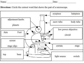
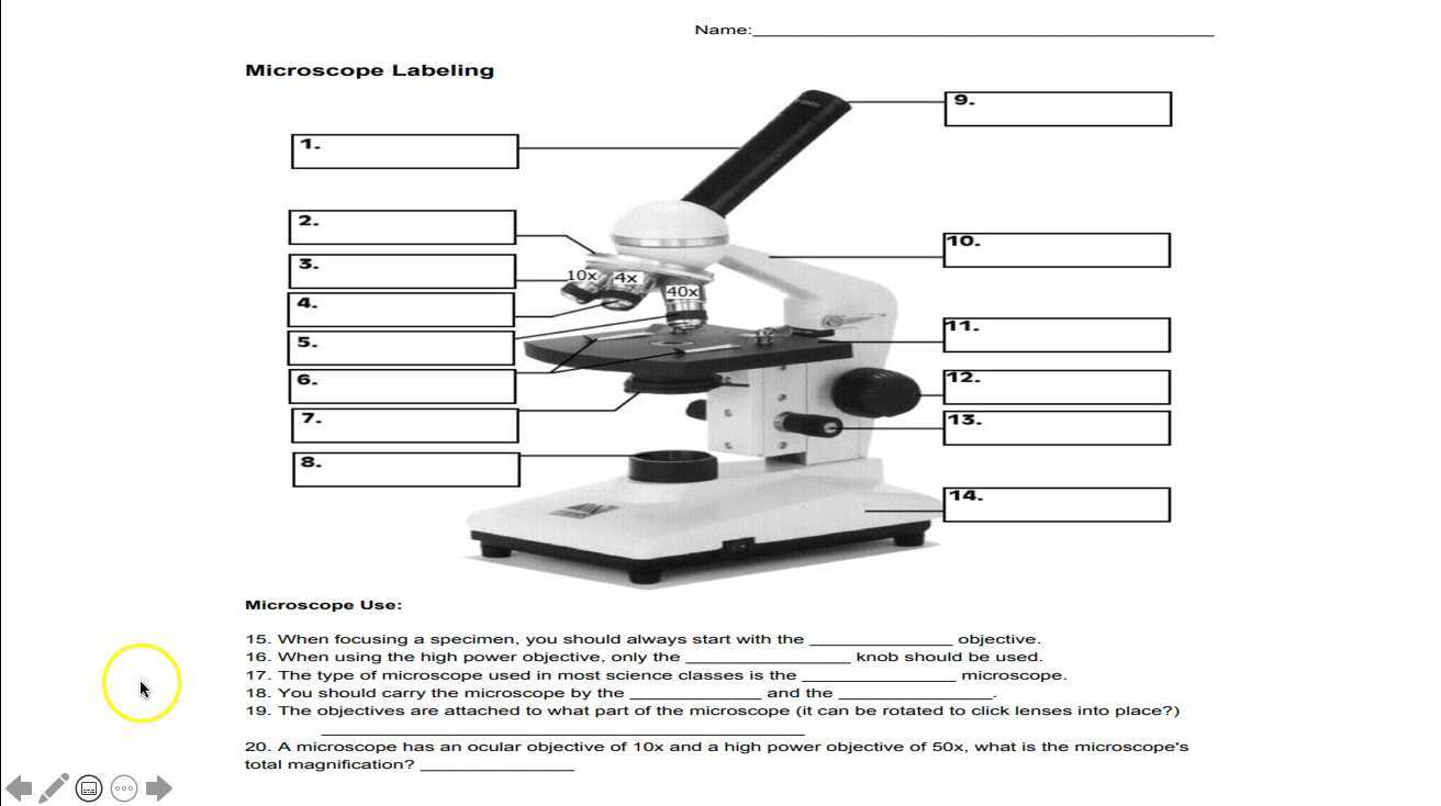
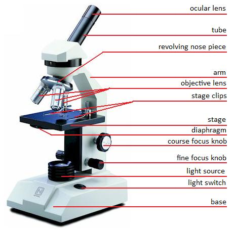
Microscope Labeling - The Biology Corner Students label the parts of the microscope in this photo of a basic laboratory light microscope. Can be used for practice or as a quiz. ... Microscope Labeling . Microscope Use: 15. When focusing a specimen, you should always start with the _____ objective. 16. When using the high power objective, only the _____ knob should be used. 17. The ...
Microscope Types (with labeled diagrams) and Functions
Labelled Diagram of Compound Microscope The below mentioned article provides a labelled diagram of compound microscope. Part # 1. The Stand: The stand is made up of a heavy foot which carries a curved inclinable limb or arm bearing the body tube. The foot is generally horse shoe-shaped structure (Fig. 2) which rests on table top or any other surface on which the microscope in kept.

Parts of a Microscope - SmartSchool Systems
Compound Microscope Parts, Functions, and Labeled Diagram Compound Microscope Definitions for Labels. Eyepiece (ocular lens) with or without Pointer: The part that is looked through at the top of the compound microscope. Eyepieces typically have a magnification between 5x & 30x. Monocular or Binocular Head: Structural support that holds & connects the eyepieces to the objective lenses.

draw a well label diagram of microscope - Brainly.in
Microscope Parts and Functions Most specimens are mounted on slides, flat rectangles of thin glass. The specimen is placed on the glass and a cover slip is placed over the specimen. This allows the slide to be easily inserted or removed from the microscope. It also allows the specimen to be labeled, transported, and stored without damage.
Microscope Label Teaching Resources | Teachers Pay Teachers
Labeling the Parts of the Microscope | Microscope World Resources Labeling the Parts of the Microscope This activity has been designed for use in homes and schools. Each microscope layout (both blank and the version with answers) are available as PDF downloads. You can view a more in-depth review of each part of the microscope here. Download the Label the Parts of the Microscope PDF printable version here.
MICROBIO 16 Parts of a Compound Microscope with Diagram and ...
Microscope Parts, Function, & Labeled Diagram - slidingmotion Microscope parts labeled diagram gives us all the information about its parts and their position in the microscope. Microscope Parts Labeled Diagram The principle of the Microscope gives you an exact reason to use it. It works on the 3 principles. Magnification Resolving Power Numerical Aperture. Parts of Microscope Head Base Arm Eyepiece Lens

Compound Microscope Parts
Welcome to Virtual Urchin - University of Washington microscope measurement. microscope compare. specimen compare. development & embryology. fertilization lab. embryogenesis to hatching. analyzing gene function. ecology & environment. our acidifying ocean. predator & prey. surfing to settlement. basic biology. urchin anatomy. about us. teacher resources. useful links . Select Language: Welcome to the new …

Labelling a Microscope Diagram | Quizlet
Label the Microscope Diagram | Download Scientific Diagram - ResearchGate Download scientific diagram | Label the Microscope Diagram from publication: Laboratory Exercises in Microbiology: Discovering the Unseen World through Hands-on Investigation | Microbiology ...

Draw a labelled diagram of a compound microscope.
A Study of the Microscope and its Functions With a Labeled Diagram ... A Study of the Microscope and its Functions With a Labeled Diagram To better understand the structure and function of a microscope, we need to take a look at the labeled microscope diagrams of the compound and electron microscope. These diagrams clearly explain the functioning of the microscopes along with their respective parts.

Simple Microscope Definition, Magnification, Parts And Uses
Parts of a Microscope Labeling Activity - Storyboard That Create a poster that labels the parts of a microscope and includes descriptions of what each part does. Click "Start Assignment". Use a landscape poster layout (large or small). Search for a diagram of a microscope. Using arrows and textables label each part of the microscope and describe its function.
simple light microscope diagram - Clip Art Library
Label the microscope — Science Learning Hub 08.06.2018 · All microscopes share features in common. In this interactive, you can label the different parts of a microscope. Use this with the Microscope parts activity to help students identify and label the main parts of a microscope and then describe their functions.. Drag and drop the text labels onto the microscope diagram. If you want to redo an answer, click on the …

Compound Microscope Parts, Diagram Definition, Application ...
Microscope, Microscope Parts, Labeled Diagram, and Functions (2022) The description given below summarize the brief description of microscope parts used to visualize the microscopic specimens such as animal cells, plant cells, microbes, bacteria, viruses, microorganisms etc. The Microscopes parts divided into three different structural parts Head, Base, and Arms.

Light Microscope- Definition, Principle, Types, Parts ...
Compound Microscope- Definition, Labeled Diagram, Principle, Parts, Uses The optical microscope often referred to as the light microscope, is a type of microscope that uses visible light and a system of lenses to magnify images of small subjects. There are two basic types of optical microscopes: Simple microscopes. Compound microscopes. The term "compound" in compound microscopes refers to the microscope having ...
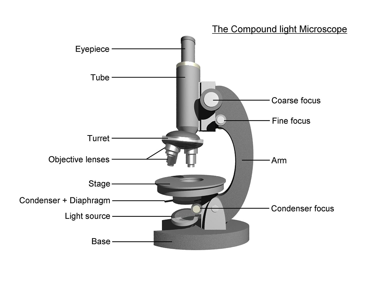
The Compound Light Microscope Label the following parts on ...
Binocular Microscope Anatomy - Parts and Functions with a Labeled Diagram The nose piece of a microscope, Head part of the microscope, Ocular lens or eyepiece of the microscope, Diopter adjustment of the eyepiece All of these parts are identified in a light microscope labeled diagram. So, first, make sure you can identify all these parts from this labeled diagram. Parts of the compound microscope
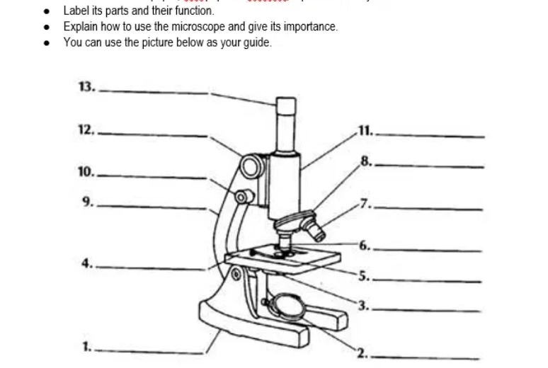
Answered: Label its parts and their function.… | bartleby
Microscope Labeling - The Biology Corner Microscope Labeling. Shannan Muskopf May 31, 2018. This simple worksheet pairs with a lesson on the light microscope, where beginning biology students learn the parts of the light microscope and the steps needed to focus a slide under high power. The labeling worksheet could be used as a quiz or as part of direct instruction where students ...

Simple Microscope - Diagram (Parts labelled), Principle ...
PDF Label parts of the Microscope: Answers Label parts of the Microscope: Answers Coarse Focus Fine Focus Eyepiece Arm Rack Stop Stage Clip . Created Date: 20150715115425Z ...

Compound Microscope- Definition, Labeled Diagram, Principle ...
Parts of a microscope with functions and labeled diagram 19.04.2022 · Figure: Diagram of parts of a microscope. There are three structural parts of the microscope i.e. head, base, and arm. Head – This is also known as the body. It carries the optical parts in the upper part of the microscope. Base – It acts as microscopes support. It also carries microscopic illuminators.

Simple Microscope - Diagram (Parts labelled), Principle ...
Fluorescence microscope - Wikipedia The majority of fluorescence microscopes, especially those used in the life sciences, are of the epifluorescence design shown in the diagram.Light of the excitation wavelength illuminates the specimen through the objective lens. The fluorescence emitted by the specimen is focused to the detector by the same objective that is used for the excitation which for greater resolution will …

Microscope, Microscope Parts, Labeled Diagram, and Functions
16 Parts of a Compound Microscope: Diagrams and Video Once you have an understanding of the parts of the microscope it will be much easier to navigate around and begin observing your specimen, which is the fun part! The 16 core parts of a compound microscope are: Head (Body) Arm. Base. Eyepiece. Eyepiece tube.

Biology - labeling a compound microscope Diagram | Quizlet
Parts of Stereo Microscope (Dissecting microscope) – labeled diagram ... Unlike a compound microscope that offers a flat image, stereo microscopes give the viewer a 3-dimensional image that you can see the texture of a larger specimen. [In this image] Examples of Stereo & Dissecting microscopes. Major microscope brands (Zeiss, Olympus, Nikon, Amscope, Omano, Leica …) all produce stereomicroscopes.

File:Microscope diagram.png - Wikimedia Commons
Microscope labeled diagram - SlideShare Microscope labeled diagram 1. The Microscope Image courtesy of: Microscopehelp.com Basic rules to using the microscope 1. You should always carry a microscope with two hands, one on the arm and the other under the base. 2. You should always start on the lowest power objective lens and should always leave the microscope on the low power lens ...
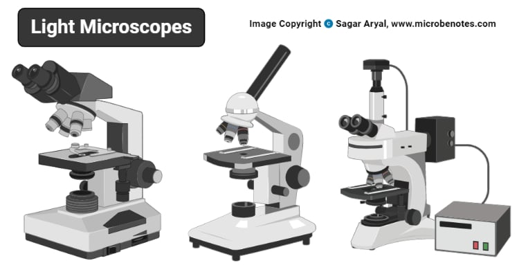
Light Microscope- Definition, Principle, Types, Parts ...
Microscope Labeling
Diagram of a Microscope - Guide to using a microscope

Microscope Diagram Labeled, Unlabeled and Blank | Parts of a ...

Microscope With Labels clip art | Microscope parts ...
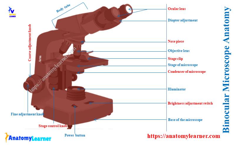
Binocular Microscope Anatomy - Parts and Functions with a ...

Microscope Diagram Labeled, Unlabeled and Blank | Parts of a ...

File:Labelledmicroscope.gif - Wikimedia Commons
Label Microscope Parts - ClipArt Best

Compound Microscope: Parts of Compound Microscope
Jual PREORDER 1600X Student Biological Microscope 2MP USB ...
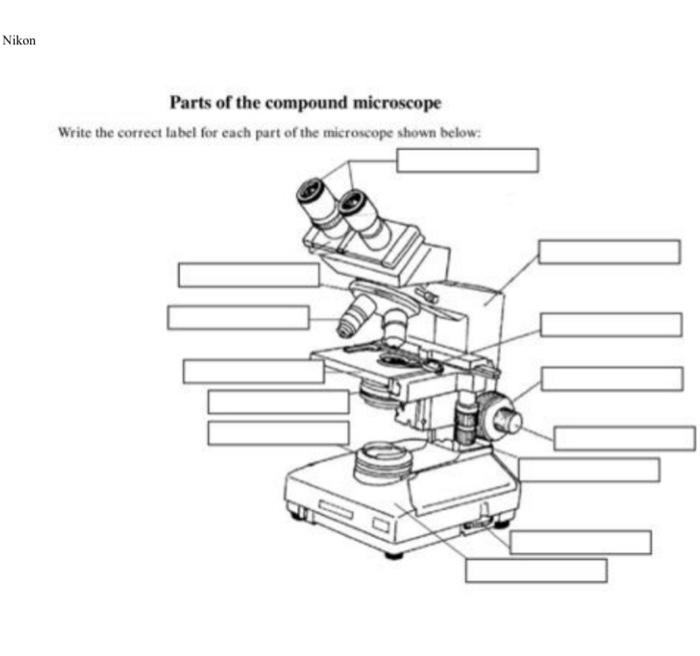
Solved Nikon Parts of the compound microscope Write the ...

Carl Zeiss Microscopy Optical microscope Worksheet Diagram ...

Compound Microscope Parts – Labeled Diagram and their ...

Microscopy: History, Types of Microscope, Diagram - Embibe
simple light microscope labeled - Clip Art Library
Parts of a Microscope Labeling Activity
Post a Comment for "38 diagram of microscope with label"