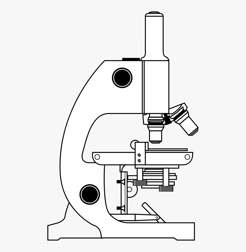
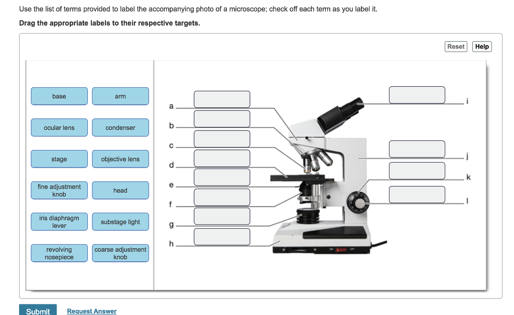
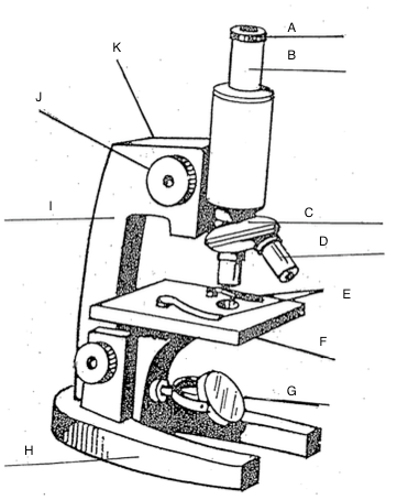
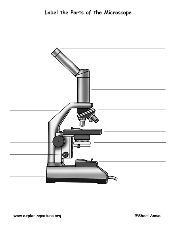
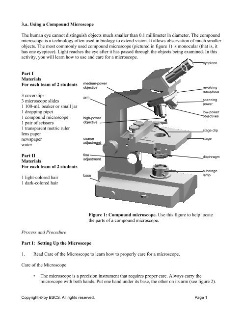
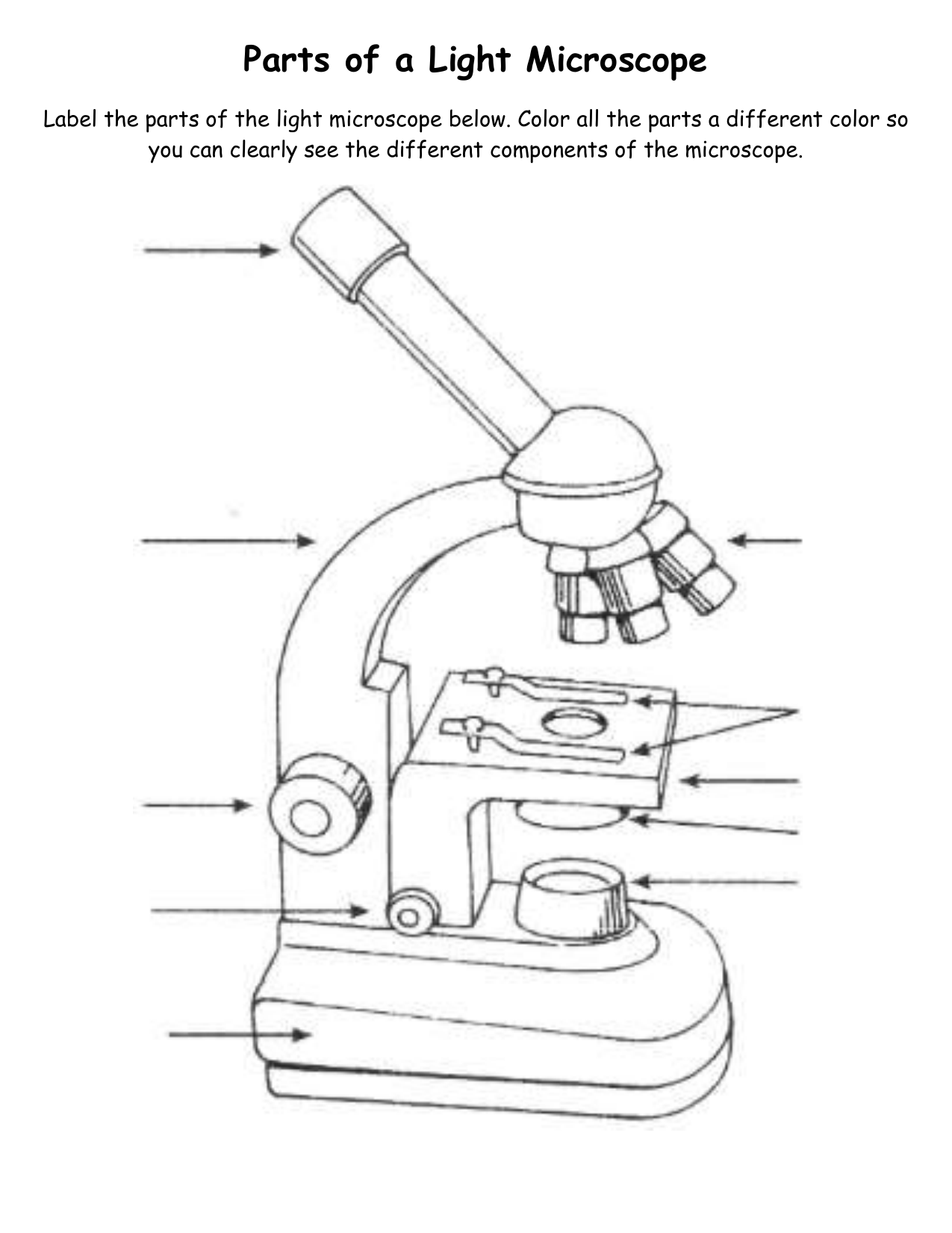
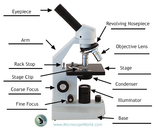
41 microscope picture with labels
› b › Sony-PlayStation-5-ConsolesSony PlayStation 5 Consoles for sale | eBay Get the best deals on Sony PlayStation 5 Consoles and upgrade your gaming setup with a new gaming console. Find the lowest prices at eBay.com. Fast & Free shipping on many items! Scanning electron microscope - Wikipedia A scanning electron microscope (SEM) is a type of electron microscope that produces images of a sample by scanning the surface with a focused beam of electrons.The electrons interact with atoms in the sample, producing various signals that contain information about the surface topography and composition of the sample. The electron beam is scanned in a raster scan …
Microscope illustrations and clipart (69,383) - Can Stock Photo Download Microscope images and photos. Over 69,383 Microscope pictures to choose from, with no signup needed. Download in under 30 seconds.

Microscope picture with labels
ZEISS Axioscan 7 Microscope Slide Scanner Digitize your specimens with Axioscan 7 – the reliable, reproducible way to create high-quality virtual microscope slides. Axioscan 7 combines qualities that you would not expect to get in a slide scanner: high speed digitization and outstanding image quality plus an unrivaled variety of imaging modes are all available in a fully automated and easy to operate system. Mitosis Images Labeled | Virtual Anatomy Lab VAL - ncccval Endocrine Rabbit Dissection Unlabeled. Cardiovascular. Cardiovascular Histology Labeled. Cardiovascular Histology Unlabeled. Cardiovascular Models Labeled. Cardiovascular Models Unlabeled. Cardiovascular Sheep Heart Dissect-L. Cardiovascular Sheep Heart Disect-U. Cardiovascular Cat Dissection Labeled. Sony PlayStation 5 Consoles for sale | eBay Get the best deals on Sony PlayStation 5 Consoles and upgrade your gaming setup with a new gaming console. Find the lowest prices at eBay.com. Fast & Free shipping on many items!
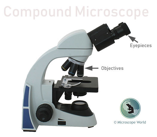
Microscope picture with labels. Module: data — skimage v0.19.2 docs - scikit-image This plant stem on a pre-prepared slide was imaged with confocal fluorescence microscopy (Nikon C1 inverted microscope). Image shape is (922, 922, 4). That is 922x922 pixels in X-Y, with 4 color channels. Real-space voxel size is 1.24 microns in X-Y. Data type is unsigned 16-bit integers. Returns lily (922, 922, 4) uint16 ndarray What is Electron Microscopy? - UMASS Medical School Conventional scanning electron microscopy depends on the emission of secondary electrons from the surface of a specimen. Because of its great depth of focus, a scanning electron microscope is the EM analog of a stereo light microscope. It provides detailed images of the surfaces of cells and whole organisms that are not possible by TEM. Compound Microscope Parts - Labeled Diagram and their Functions There are two major optical lens parts of a microscope: Eyepiece (10x) and Objective lenses (4x, 10x, 40x, 100x). Total magnification power is calculated by multiplying the magnification of the eyepiece and objective lens. The illuminator provides a source of light. The light is focused by the condenser and passing through the specimen placed ... Microscope Labeled Pictures, Images and Stock Photos Browse 49 microscope labeled stock photos and images available, or start a new search to explore more stock photos and images. Newest results.
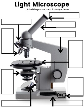
Microscope picture label Flashcards | Quizlet Start studying Microscope picture label. Learn vocabulary, terms, and more with flashcards, games, and other study tools. Microscope Images Labeled | Virtual Anatomy Lab VAL - ncccval Body cavities, planes, and regions. Body Images Labeled. Body Images Unlabeled. Histology. Epithelium Images Labeled. Epithelium Images Unlabeled. Connective Tissue Images Labeled. Connective Tissue Images Unlabeled. Microscope. Electron Microscopy Images - Dartmouth Transmission electron microscope image of a thin section cut through the bronchiolar epithelium of the lung (mouse), which consists of ciliated cells and non-ciliated cells. Image shows the ciliary microtubules in transverse and oblique section. In the cell apex are the basal bodies that are the anchoring sites for the cilia. Labeling the Parts of the Microscope | Microscope World Resources Labeling the Parts of the Microscope. This activity has been designed for use in homes and schools. Each microscope layout (both blank and the version with answers) are available as PDF downloads. You can view a more in-depth review of each part of the microscope here.
scikit-image.org › docs › stableModule: data — skimage v0.19.2 docs - scikit-image This plant stem on a pre-prepared slide was imaged with confocal fluorescence microscopy (Nikon C1 inverted microscope). Image shape is (922, 922, 4). That is 922x922 pixels in X-Y, with 4 color channels. Real-space voxel size is 1.24 microns in X-Y. Data type is unsigned 16-bit integers. Returns lily (922, 922, 4) uint16 ndarray Microscope Types (with labeled diagrams) and Functions A compound microscope: Is used to view samples that are not visible to the naked eye. Uses two types of lenses - Objective and ocular lenses. Has a higher level of magnification - Typically up to 2000x. Is used in hospitals and forensic labs by scientists, biologists and researchers to study micro organisms. › microscopy › enZEISS Axioscan 7 Microscope Slide Scanner Digitize your specimens with Axioscan 7 – the reliable, reproducible way to create high-quality virtual microscope slides. Axioscan 7 combines qualities that you would not expect to get in a slide scanner: high speed digitization and outstanding image quality plus an unrivaled variety of imaging modes are all available in a fully automated and easy to operate system. 300+ Free Microscope & Laboratory Images - Pixabay 399 Free images of Microscope. Related Images: laboratory science bacteria research scientist lab biology chemistry medical. Find your perfect microscope image. Free pictures to download and use in your next project. 399 Free images of Microscope / 4 ‹ › ...
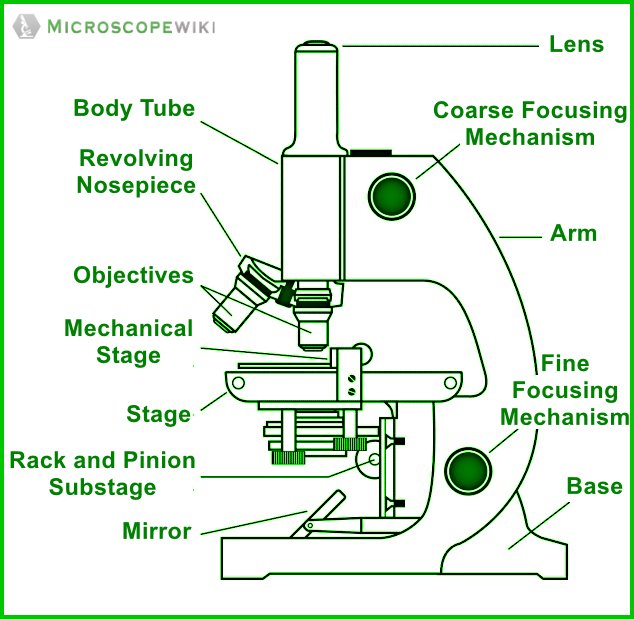
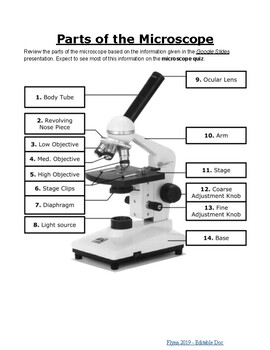
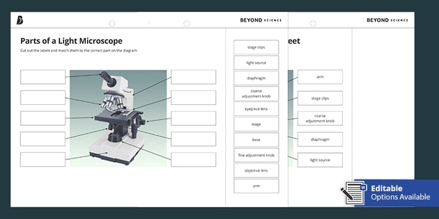
Parts of a microscope with functions and labeled diagram - Microbe Notes Head - This is also known as the body. It carries the optical parts in the upper part of the microscope. Base - It acts as microscopes support. It also carries microscopic illuminators. Arms - This is the part connecting the base and to the head and the eyepiece tube to the base of the microscope.
› cemf › whatisemWhat is Electron Microscopy? - UMASS Medical School Conventional scanning electron microscopy depends on the emission of secondary electrons from the surface of a specimen. Because of its great depth of focus, a scanning electron microscope is the EM analog of a stereo light microscope. It provides detailed images of the surfaces of cells and whole organisms that are not possible by TEM.
Label the microscope — Science Learning Hub All microscopes share features in common. In this interactive, you can label the different parts ...
recorder.butlercountyohio.org › search_records › subdivisionWelcome to Butler County Recorders Office Copy and paste this code into your website. Your Link Name
Labelled Diagram of Compound Microscope - Biology Discussion The below mentioned article provides a labelled diagram of compound microscope. Part # 1. The Stand: The stand is made up of a heavy foot which carries a curved inclinable limb or arm bearing the body tube. The foot is generally horse shoe-shaped structure (Fig. 2) which rests on table top or any other surface on which the microscope in kept.
Parts of the Microscope with Labeling (also Free Printouts) 5. Knobs (fine and coarse) By adjusting the knob, you can adjust the focus of the microscope. The majority of the microscope models today have the knobs mounted on the same part of the device. Image 5: The circled parts of the microscope are the fine and coarse adjustment knobs. Picture Source: bp.blogspot.com.
Welcome to Butler County Recorders Office Copy and paste this code into your website. Your Link …
The Parts of a Microscope (Labeled) Printable - TeacherVision The Parts of a Microscope (Labeled) Printable. Download. Add to Favorites. Share. This diagram labels and explains the function of each part of a microscope. Use this printable as a handout or transparency to help prepare students for working with laboratory equipment.
PDF Label parts of the Microscope Label parts of the Microscope: . Created Date: 20150715115425Z
Looking at the Structure of Cells in the Microscope A typical animal cell is 10–20 μm in diameter, which is about one-fifth the size of the smallest particle visible to the naked eye. It was not until good light microscopes became available in the early part of the nineteenth century that all plant and animal tissues were discovered to be aggregates of individual cells. This discovery, proposed as the cell doctrine by Schleiden and …
› books › NBK26880Looking at the Structure of Cells in the Microscope ... The phase-contrast microscope and, in a more complex way, the differential-interference-contrast microscope, exploit the interference effects produced when these two sets of waves recombine, thereby creating an image of the cell's structure . Both types of light microscopy are widely used to visualize living cells.
Microscope Parts and Functions Microscope Parts and Functions With Labeled Diagram and Functions How does a Compound Microscope Work?. Before exploring microscope parts and functions, you should probably understand that the compound light microscope is more complicated than just a microscope with more than one lens.. First, the purpose of a microscope is to magnify a small object or to magnify the fine details of a larger ...
microscope picture with labels - Compound Light Microscope... View microscope picture with labels from BIOL 1005Y at Yeshiva University. Compound Light Microscope ocular (eyepiece) revolving nosepiece objectives coarse adjustment knob mechanical stage fine
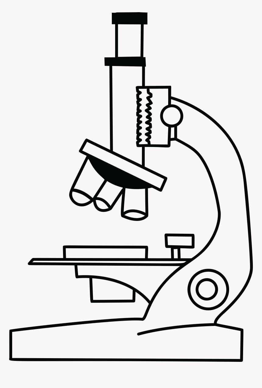
Drawing Of A Microscope And Label - Warehouse of Ideas The optical parts of the microscope are used to view, magnify, and produce an image from a specimen placed on a slide. Source: microspedia.blogspot.com. Learn how to draw microscope and label pictures using these outlines or print just for coloring. Controls the amount of light passing through the opening of the stage.
Microscope Parts, Function, & Labeled Diagram - slidingmotion Objective lenses. Objective lenses are the most important part of the microscope. Its purpose is to visualize the specimen. There are 3-4 types of different objective lenses in any microscope. It has a magnification power of 4X to 100 X. 4X objective lens is the shortest lens while the 100X lens is the longest in terms of visualization.
Assignment Essays - Best Custom Writing Services Get 24⁄7 customer support help when you place a homework help service order with us. We will guide you on how to place your essay help, proofreading and editing your draft – fixing the grammar, spelling, or formatting of your paper easily and cheaply.
en.wikipedia.org › wiki › Scanning_electron_microscopeScanning electron microscope - Wikipedia History. An account of the early history of scanning electron microscopy has been presented by McMullan. Although Max Knoll produced a photo with a 50 mm object-field-width showing channeling contrast by the use of an electron beam scanner, it was Manfred von Ardenne who in 1937 invented a microscope with high resolution by scanning a very small raster with a demagnified and finely focused ...
Amazon.com : Celestron 44302 Deluxe Handheld Digital USB Microscope … The intermediate-level Celestron Deluxe Handheld Digital Microscope is an easy to use, low power microscope. With powers of 10x to 40x, it’s ideal for viewing stamps, coins, bugs, plants, rocks, skin, gems, circuit boards, and more. With the higher power magnifications, you can even view traditional microscope slides.
Home: The Histology Guide You can see histological slides on the pages and can turn labels on or off to help them identify features. In some cases, there is a section like a 'virtual microscope' - you can scan around a large picture using the mouse and try to identify features. This emulates as closely as possible the experience of using a microscope.
Sony PlayStation 5 Consoles for sale | eBay Get the best deals on Sony PlayStation 5 Consoles and upgrade your gaming setup with a new gaming console. Find the lowest prices at eBay.com. Fast & Free shipping on many items!
Mitosis Images Labeled | Virtual Anatomy Lab VAL - ncccval Endocrine Rabbit Dissection Unlabeled. Cardiovascular. Cardiovascular Histology Labeled. Cardiovascular Histology Unlabeled. Cardiovascular Models Labeled. Cardiovascular Models Unlabeled. Cardiovascular Sheep Heart Dissect-L. Cardiovascular Sheep Heart Disect-U. Cardiovascular Cat Dissection Labeled.
ZEISS Axioscan 7 Microscope Slide Scanner Digitize your specimens with Axioscan 7 – the reliable, reproducible way to create high-quality virtual microscope slides. Axioscan 7 combines qualities that you would not expect to get in a slide scanner: high speed digitization and outstanding image quality plus an unrivaled variety of imaging modes are all available in a fully automated and easy to operate system.














Post a Comment for "41 microscope picture with labels"